
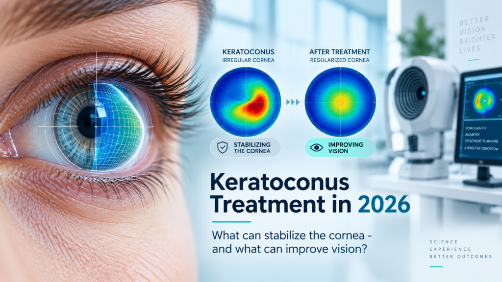
One of the most confusing things about keratoconus treatment is that two very different goals are often discussed as if they were the same.
The first goal is to stop or slow the cornea from becoming more ectatic.
The second goal is to help the patient see better.
A treatment that stabilizes keratoconus does not necessarily restore clear vision. Likewise, a contact lens that provides excellent vision does not stop the underlying cornea from progressing.
Understanding this distinction is one of the most important parts of modern keratoconus management.
For a broader overview of the condition, diagnosis and specialty lens options, visit the main Keratoconus treatment and diagnosis page.
Goal 1 – Stabilizing the cornea
When keratoconus is progressing, the medical priority is often to reduce the risk of further structural deterioration.
Corneal collagen cross-linking, or CXL, is designed for this purpose. Riboflavin and ultraviolet-A light are used to increase biomechanical stability within the corneal tissue.
The key word is “stability”.
CXL is not primarily a vision-correction procedure. Some patients may experience changes in corneal shape or vision after treatment, but its central therapeutic purpose is to reduce progression.
This distinction is particularly important for younger patients and for anyone whose corneal maps show measurable progression over time.
What changed by 2026?
Cross-linking itself is not new, but the treatment landscape continues to evolve.
In the United States, the FDA had already approved an epithelium-off CXL system for progressive keratoconus in 2016. A major newer development came in October 2025, when the FDA approved Epioxa and Epioxa HD for epithelium-on corneal collagen cross-linking in patients aged 13 years and older, used with the designated treatment system.
That makes 2026 an important point in the evolution of CXL because epithelium-on treatment is no longer only an investigational concept in the United States.
However, regulatory approval and availability differ between countries. Patients in Israel, the UAE, Saudi Arabia, Qatar, Bahrain, Kuwait, Turkey and other markets should ask their ophthalmologist which CXL protocols are locally approved, available and appropriate for their individual corneal thickness and progression pattern.
Epi-off versus epi-on does not replace the need for diagnosis
A newer protocol does not mean every patient should receive it.
The decision to perform cross-linking depends on factors such as documented progression, age, corneal thickness, topography or tomography findings and the overall condition of the cornea.
In 2026, the conversation is becoming more personalized, not less.
Research also continues into customized cross-linking patterns. A recent meta-analysis found that customized CXL can produce different topographic outcomes from non-customized protocols, but patient selection and long-term evidence remain important.
Goal 2 – Improving vision
Once stability has been addressed, a different question remains:
“How can I actually see better?”
In early keratoconus, glasses or selected soft contact lenses may provide adequate vision.
As the cornea becomes more irregular, standard correction often becomes less effective because glasses correct conventional refractive error but cannot fully neutralize complex corneal distortion.
This is where specialty contact lenses become important.
RGP, hybrid and scleral lenses
Different lens designs can be used depending on the shape of the cornea, the severity of the ectasia, ocular surface condition and patient tolerance.
Scleral lenses have become particularly important in moderate and advanced keratoconus because they vault over the irregular cornea and create a new optical surface.
They do not reverse keratoconus and they do not replace cross-linking when the disease is progressing.
Their role is visual rehabilitation.
Our detailed guide to scleral lenses for keratoconus – advantages, disadvantages and fitting considerations explains this side of treatment in greater depth.
Recent literature continues to show major advances in scleral lens fitting, including customized haptics, profilometry, impression-based designs and wavefront-guided optics for highly irregular eyes.
Why 20/20 is not always the final goal
Some keratoconus patients achieve acceptable visual acuity with a specialty lens but still complain about halos, glare, starbursts, ghost images or poor night vision.
This can happen because keratoconus creates Higher-Order Aberrations, or HOA, that may remain even after standard refractive correction.
For selected complex cases, advanced scleral lens optics can be designed to address measured higher-order aberrations.
This is a different level of treatment from simply correcting sphere and cylinder.
Patients experiencing this type of visual problem can read more about HOA-correcting scleral lenses and advanced wavefront optics.
Does cross-linking make specialty lenses unnecessary?
No.
This is one of the biggest misconceptions in keratoconus.
A successful cross-linking procedure can stabilize the cornea while the patient still has irregular astigmatism and poor visual quality.
That patient may continue to need glasses, RGP lenses, hybrid lenses or scleral lenses.
In simple terms:
Cross-linking addresses progression.
Specialty optics address vision.
Many patients need both strategies at different points in their care.
For patients concerned about severe progression, our article Does Keratoconus Lead to Blindness Without Treatment? explains what advanced disease can mean and why early management matters.
What about corneal rings and newer procedures?
Intrastromal corneal ring segments have been used in selected keratoconus cases to modify corneal shape.
Another developing approach involves Corneal Allogenic Intrastromal Ring Segments, commonly called CAIRS, which use donor corneal tissue rather than synthetic ring material.
A 2025 systematic review and meta-analysis reported promising visual and topographic outcomes for CAIRS, but this remains a specialized surgical approach and should not be presented as a universal replacement for CXL or specialty lenses.
The correct procedure depends on the individual cornea and must be evaluated by a corneal ophthalmologist.
What about laser treatment combined with cross-linking?
This is another area that receives a lot of attention online.
Selected centers investigate combinations of topography-guided or wavefront-guided PRK with CXL, aiming to improve corneal regularity while also addressing progression.
But this is not the same as routine laser vision correction in a normal cornea.
As of 2026, clinical research is still actively comparing different combined strategies. For example, an ongoing randomized trial is comparing corneal wavefront-guided PRK plus epi-off CXL with another CXL-based approach in keratoconus.
The takeaway is not that “laser now cures keratoconus”. The takeaway is that treatment is becoming increasingly customized.
When is a corneal transplant still necessary?
Corneal transplantation remains relevant for selected advanced cases, particularly when severe scarring, extreme thinning or structural changes prevent useful vision with other approaches.
But a transplant does not necessarily eliminate the need for optical rehabilitation afterward.
Patients may still have irregular astigmatism and can require specialty lenses after surgery.
For a deeper discussion of complex disease, see Advanced Keratoconus and Corneal Diseases – When Standard Solutions Are No Longer Enough.
How should treatment decisions be made in 2026?
The most useful way to think about keratoconus is not:
“What is the best treatment?”
Instead, ask four separate questions.
Is the cornea progressing?
Does it need medical stabilization?
What is currently limiting the patient’s vision?
Which optical solution provides the best usable vision and comfort?
The answers may lead to completely different interventions.
An ophthalmologist may manage progression and surgical treatment, while an optometrist experienced in irregular corneas can focus on visual rehabilitation, advanced measurements and specialty lens fitting.
Keratoconus care at M’Eye Clinic in Jerusalem
M’Eye Clinic in Jerusalem, Israel works with patients whose keratoconus affects real-world visual quality and cannot be adequately corrected with standard glasses or conventional contact lenses.
The clinic provides advanced evaluation and customized optical rehabilitation using a wide range of specialty contact lens technologies, including scleral lenses, customized designs, EyePrintPRO and advanced HOA-related optical strategies where appropriate.
Patients also travel to Jerusalem from countries across the Middle East, including the UAE, Saudi Arabia, Qatar, Bahrain and Kuwait, when they require evaluation of complex corneal and specialty lens cases.
FAQ
Can keratoconus be cured in 2026?
There is still no single treatment that simply restores a keratoconic cornea to a completely normal cornea. Modern management aims to stabilize progression and achieve the best possible visual rehabilitation.
Does cross-linking improve vision?
Its main purpose is stabilization. Some patients experience optical changes after treatment, but vision correction may still be required.
Do scleral lenses stop keratoconus from progressing?
No. Scleral lenses are primarily used to improve visual quality and, in selected cases, comfort. Progression must be evaluated separately.
Is epi-on cross-linking now approved?
In the United States, an epithelium-on CXL system received FDA approval in October 2025 for keratoconus in patients aged 13 years and older. Availability and regulatory status differ by country.
Are newer treatments replacing scleral lenses?
No. New surgical and corneal stabilization techniques address different goals. A patient can have a successfully stabilized cornea and still benefit greatly from advanced specialty lenses.
Final thought
The biggest advance in keratoconus treatment is not one single device or procedure.
It is the shift toward treating the disease according to its actual objective.
First determine whether the cornea is progressing.
Then decide whether it needs stabilization.
Then determine what is limiting vision.
Finally, choose the optical rehabilitation that gives the patient the clearest, most stable and most functional vision possible.
In 2026, keratoconus care is increasingly about combining these strategies rather than expecting one treatment to do everything.